The female reproductive system is a network of internal and external organs responsible for menstruation, reproductive hormone production, sexual function, fertility, pregnancy, and childbirth.
Its major structures include the ovaries, fallopian tubes, uterus, cervix, vagina, and vulva. Each performs a distinct function, but reproductive processes depend on their coordinated activity.
The ovaries develop and release eggs, the fallopian tubes provide the usual site of fertilization, and the uterus supports implantation and pregnancy. Hormonal signals from the brain and ovaries regulate these processes.
Understanding reproductive anatomy also helps explain menstrual cycles, hormonal changes, and common reproductive health concerns.
This guide covers the major organs, their locations and functions, the menstrual cycle, fertilization, pregnancy, and changes throughout life.
What Is the Female Reproductive System?
The female reproductive system consists of specialized organs and tissues that support reproductive and sexual functions.
It has several important roles:
- Developing and releasing egg cells.
- Producing reproductive hormones.
- Regulating menstrual cycles.
- Providing pathways for sperm and eggs.
- Supporting fertilization and implantation.
- Maintaining pregnancy and facilitating childbirth.
The reproductive system works closely with the endocrine system, particularly the hypothalamus and pituitary gland.
Its anatomy and function can vary naturally between individuals. Some people are born with anatomical differences or undergo medical procedures that alter reproductive structures.
Female Reproductive System Anatomy: Main Parts and Locations
The reproductive organs are generally divided into internal and external structures.
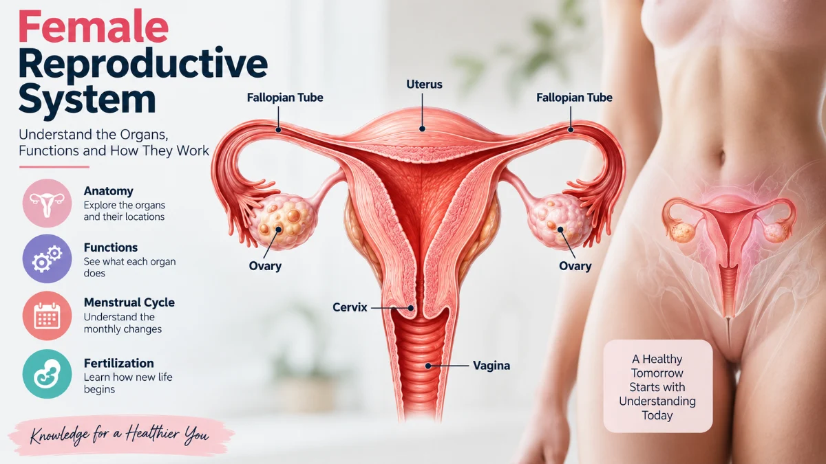
Female reproductive system anatomy: the ovaries, fallopian tubes, uterus, cervix, and vagina.
Internal reproductive organs
| Organ | Location | Primary function |
| Ovaries | Either side of the uterus | Egg development and hormone production |
| Fallopian tubes | Extend from the uterus toward the ovaries | Egg transport and usual site of fertilization |
| Uterus | Pelvic cavity | Menstruation and pregnancy support |
| Cervix | Lower portion of the uterus | Connects the uterus and vagina |
| Vagina | Between the cervix and vaginal opening | Passage for menstrual flow and vaginal childbirth |
These structures coordinate to support reproductive processes.
Read More: Ocular Migraine
External reproductive organs
The external genital structures are collectively called the vulva.
They include the labia majora, labia minora, clitoris, mons pubis, vaginal opening, and surrounding tissues.
The urethral opening is located in this region but belongs to the urinary system.
The vulva protects sensitive structures and contributes to sexual sensation.
Internal Female Reproductive Organs Explained
1. Ovaries
The ovaries are small, oval-shaped glands located on either side of the uterus.
They perform two major functions: developing eggs and producing hormones.
Each ovary contains follicles that hold immature egg cells. During a typical ovulatory menstrual cycle, several follicles begin developing, but usually one becomes dominant.
The dominant follicle eventually releases an egg through ovulation.
The ovaries also produce estrogen and progesterone, which influence the menstrual cycle and reproductive tissues.
Ovarian activity changes throughout life and generally declines as menopause approaches.
2. Fallopian Tubes
The fallopian tubes are narrow structures extending from the upper uterus toward the ovaries.
Their principal function is transporting an egg toward the uterus.
Finger-like projections called fimbriae help capture the egg after ovulation.
Fertilization usually occurs in the ampulla, a wider section of the fallopian tube.
Cilia and muscular contractions help transport the egg or developing embryo toward the uterus.
The fallopian tubes therefore play an essential role in natural conception.
3. Uterus
The uterus is a hollow, muscular organ situated in the pelvis between the bladder and rectum.
It is commonly described as pear-shaped, although its position and dimensions vary.
The uterine wall contains three major layers.
| Layer | Function |
| Endometrium | Changes during the menstrual cycle and supports implantation |
| Myometrium | Muscular layer responsible for contractions |
| Perimetrium | Outer covering of the uterus |
During the menstrual cycle, the endometrium develops in response to reproductive hormones.
If implantation does not occur, the lining is shed during menstruation.
During pregnancy, the uterus expands to accommodate fetal development. Its muscular contractions also contribute to childbirth.
4. Cervix
The cervix forms the lower part of the uterus and connects it to the vagina.
Its narrow canal allows menstrual blood to leave the uterus and provides a passage for sperm.
Cervical mucus changes throughout the menstrual cycle.
Around ovulation, it generally becomes thinner and more favorable for sperm movement.
During pregnancy, the cervix helps maintain the uterine environment.
During labor, it softens, thins, and dilates to facilitate vaginal childbirth.
5. Vagina
The vagina is a flexible muscular canal extending from the cervix to the vaginal opening.
Its functions include allowing menstrual blood to leave the body, receiving a penis during vaginal intercourse, and serving as the birth canal during vaginal delivery.
The vagina also contains a microbial community that contributes to its normal environment.
It is important to distinguish the vagina from the vulva.
The vagina is an internal canal, whereas the vulva refers to the external genital structures.
External Anatomy: Understanding the Vulva
The vulva consists of several structures with protective and sensory functions.
Mons pubis: A rounded area of fatty tissue located over the pubic bone.
Labia majora: Outer skin folds that protect the more delicate external structures.
Labia minora: Inner skin folds surrounding the vestibule.
Clitoris: An organ containing extensive nerve endings and erectile tissue that contributes to sexual pleasure.
Vaginal opening: The external entrance to the vagina.
Greater vestibular glands: Also called Bartholin glands, these produce secretions near the vaginal opening.
Vulvar anatomy varies naturally in size, shape, color, and appearance.
What Are the Main Functions of the Female Reproductive System?
The reproductive organs work together to perform several biological functions.
Egg development and ovulation
The ovaries support egg development and release an egg during ovulation in a typical ovulatory cycle.
Hormone production
The ovaries produce estrogen and progesterone, which regulate reproductive tissues and menstrual activity.
Menstruation
Hormonal changes cause the uterine lining to develop and, when pregnancy does not occur, shed.
Fertilization
The reproductive tract provides a pathway through which sperm can reach an egg.
Pregnancy
The uterus provides the environment for implantation and fetal development.
Childbirth
Uterine contractions and cervical dilation help facilitate vaginal delivery.
For a detailed explanation of these processes, see the supporting article on female reproductive system functions.
How Does the Menstrual Cycle Work?
The menstrual cycle involves coordinated hormonal changes in the ovaries and uterus.
It is measured from the first day of one menstrual period to the first day of the next.
According to the American College of Obstetricians and Gynecologists, most adult menstrual cycles last 21–35 days. Adolescent cycles can be longer and more variable.
Menstrual phase
Menstrual bleeding begins when declining hormone levels cause the uterine lining to shed.
Follicular phase
Follicle-stimulating hormone encourages ovarian follicles to develop.
Increasing estrogen stimulates the uterine lining to rebuild.
Ovulation
A surge in luteinizing hormone triggers the release of an egg from a mature ovarian follicle.
Ovulation timing varies and should not automatically be assumed to occur on day 14.
Luteal phase
After ovulation, the remaining follicle becomes the corpus luteum.
It produces progesterone, which prepares the uterine lining for possible implantation.
If pregnancy does not occur, hormone levels decline and menstruation begins again.
Hormones That Regulate the Reproductive System
Four major hormones coordinate reproductive cycling.
| Hormone | Main role |
| Estrogen | Supports reproductive tissue development and endometrial growth |
| Progesterone | Prepares the uterine lining for implantation |
| FSH | Stimulates ovarian follicle development |
| LH | Triggers ovulation and supports corpus luteum formation |
Hormonal regulation begins in the hypothalamus, which releases gonadotropin-releasing hormone.
This stimulates the pituitary gland to release FSH and LH.
These hormones act on the ovaries, influencing estrogen and progesterone production.
The hormones then regulate changes in the uterus and provide feedback to the brain.
How Fertilization and Pregnancy Occur
Fertilization occurs when a sperm cell joins with an egg, usually inside a fallopian tube.
The resulting cell is called a zygote.
As it travels toward the uterus, it divides repeatedly and develops into an early embryo.
A developing blastocyst may then implant into the uterine lining.
Following implantation, pregnancy-related hormonal changes support continued development.
The placenta subsequently develops and facilitates the exchange of oxygen, nutrients, and metabolic waste between maternal and fetal circulation.
Fertilization alone does not guarantee successful implantation or an ongoing pregnancy.
How the Reproductive System Changes Throughout Life
Puberty
Hormonal activation causes reproductive organs to mature and menstrual periods to begin.
Reproductive years
Ovulation and menstrual cycles generally occur, although cycle patterns vary.
Pregnancy
Hormonal and anatomical changes support embryo and fetal development.
Perimenopause
Ovarian hormone production becomes more variable, and menstrual patterns may change.
Menopause
Menopause marks the permanent end of natural menstrual periods.
It is generally identified retrospectively after 12 consecutive months without menstruation when no other cause explains the absence of periods.
Common Conditions Affecting Reproductive Organs
Several medical conditions can affect reproductive anatomy and function.
Polycystic ovary syndrome: A hormonal and metabolic condition that can interfere with regular ovulation.
Endometriosis: A condition in which tissue similar to the uterine lining develops outside the uterus.
Uterine fibroids: Generally benign growths arising from uterine muscle tissue.
Ovarian cysts: Fluid-filled structures that may develop within or on an ovary.
Pelvic inflammatory disease: Infection and inflammation affecting the upper reproductive tract.
Symptoms and treatment requirements vary considerably.
For detailed information about disorders and preventive care, read the supporting article on female reproductive system health.
When Should You See a Doctor?
Consult a healthcare professional if you experience persistent pelvic pain, unusually heavy menstrual bleeding, bleeding between periods, bleeding after menopause, persistent abnormal vaginal discharge, pain during intercourse, unexplained menstrual irregularity, or difficulty becoming pregnant.
Sudden severe pelvic pain, particularly with dizziness, fainting, or possible pregnancy, requires urgent medical evaluation.
A clinician can recommend appropriate examinations or investigations based on individual symptoms.
Frequently Asked Questions
What are the five main internal female reproductive organs?
The ovaries, fallopian tubes, uterus, cervix, and vagina are the major internal structures commonly described in reproductive anatomy.
What is the primary function of the female reproductive system?
Its functions include hormone production, egg development, menstruation, sexual function, fertilization, pregnancy, and childbirth.
Where does fertilization usually occur?
Fertilization usually occurs in the ampulla of a fallopian tube.
What is the difference between the vagina and vulva?
The vagina is an internal muscular canal. The vulva consists of the external genital structures.
Which hormones regulate the menstrual cycle?
Estrogen, progesterone, follicle-stimulating hormone, and luteinizing hormone are major regulators.
Does every menstrual cycle include ovulation?
No. Some cycles are anovulatory, meaning an egg is not released.